

EXPLORING THE DEPTHS: A Practical Manual on Industrial and Metrological Tomography

Pubblicato da Brigida Michele in Xrayconsult · Martedì 26 Mar 2024
Tags: BLOG, ESPLORANDO, LE, PROFONDITÀ, Tomografia, Industriale, Metrologica, Qualità, Affidabilità, Materiali, Prodotti, Raggi, X, Elaborazione, Digitale, Immagini, Strutture, Intricate, Dettagli, Minuziosi, Visibilità.
Tags: BLOG, ESPLORANDO, LE, PROFONDITÀ, Tomografia, Industriale, Metrologica, Qualità, Affidabilità, Materiali, Prodotti, Raggi, X, Elaborazione, Digitale, Immagini, Strutture, Intricate, Dettagli, Minuziosi, Visibilità.
EXPLORING THE DEPTHS:
A Practical Manual on Industrial and Metrological Tomography
Chapter 1: "Peeking Inside: An Introduction to Industrial Tomography"Chapter 2: "Penetrating the Veil: Fundamentals of Metrological Tomography"Chapter 3: Exploring the Different Tomography Systems: Mini-focus, Micro-focus, and Nano-focusChapter 4: "Beyond the Image: Processing and Analysis of Tomographic Data"Chapter 5: "Industrial Applications: From Automotive to Electronic Components"Chapter 6: "Navigating Anomalies: Detection and Analysis of Defects"Chapter 7: "Enhancing Precision: Precision Metrology with Tomography"Chapter 8: "Metrological Tomography vs Traditional Tomography: Key Differences in Application and Purpose"Chapter 9: "Challenges and Solutions: Optimizing Tomographic Processes"Chapter 10: "Looking to the Future: Trends and Developments in the Tomography Industry"Chapter 11: "Ethical Nuances: Considerations on the Use of Industrial Tomography"Chapter 12: "Integrated Methods: Combining Tomography with Other Inspection and Measurement Techniques"
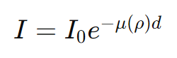
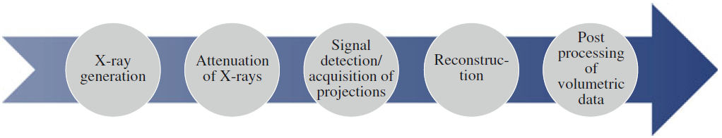
Introduction:Welcome to a journey into the unknown, illuminated by the powerful light of industrial and metrological tomography.In a world where superficial observation often masks the underlying complexity, these technologies offer us an unprecedented window to explore and understand what lies beyond the surface.Through non-invasive inspection and precision measurement, industrial and metrological tomography lead us into a world of hidden details and revealed secrets.Throughout this article, we will dive deep into the depths of tomography, examining its fundamental principles, its applications across various industrial sectors, and its implications for the future of engineering and science.From its inception as a diagnostic tool in the medical field to its evolution as a crucial technology for the non-destructive inspection of industrial components and materials, tomography has established itself as one of the most powerful and versatile imaging techniques available today.Understanding industrial and metrological tomography opens up a world of possibilities, where the quality and reliability of materials and products can be assessed with unprecedented precision.What was once invisible becomes tangible through the magic of X-rays and digital image processing, leading us to explore intricate structures and meticulous details that would otherwise remain hidden from our view.Through this article, we will venture into the heart of tomography, exploring its practical applications, technological challenges, and future development possibilities.With the goal of providing a comprehensive and accessible overview of this fascinating discipline, we hope to inspire and inform anyone interested in fully understanding and leveraging the potential of industrial and metrological tomography."Overview of a Tomographic Analysis Process"Chapter 1: "Peeking Inside: An Introduction to Industrial Tomography"In this chapter, we will dive into the basics of industrial tomography, exploring its fundamental principles, imaging techniques, and its applications across various industrial sectors.We will discover how this technology allows us to "peek" inside objects without damaging them, revolutionizing the inspection process.Peeking Inside: An Introduction to Industrial TomographyIndustrial tomography stands as one of the most revolutionary innovations in the field of non-destructive inspection.This technology enables the acquisition of three-dimensional images of objects without the need to damage them, providing a detailed view of their internal structure."Dimensional Analysis - Porosity Analysis - Deviation Analysis from CAD Design"Advanced Principles and Applications of Industrial Tomography with a Quantitative FocusIndustrial tomography utilizes advanced techniques for the non-destructive analysis of an object's internal structure by leveraging the principle of differential radiation absorption, such as X-rays or gamma rays.This methodology relies on the unique property of materials to attenuate radiation to varying degrees, allowing for a detailed examination of the internal composition without altering the object.Acquisition and Reconstruction ProcessWhen an object is exposed to a radiation source and rotated, the rays pass through the object from various angles, being captured by a detector.The attenuation of the rays, I, depends on the density, ρ, and the thickness, d, of the material traversed, following the law of exponential absorption:where I0 is the initial intensity of the rays before entering the object, and μ(ρ) is the mass attenuation coefficient, which depends on the material's density.By collecting radiographic projections from multiple angles, it's possible to apply algorithms for filtered back projection or iterative reconstruction to calculate the three-dimensional density distribution within the object.This process results in detailed three-dimensional images, or tomograms, revealing the internal structure.Applications and Benefits Industrial tomography is applied across various critical sectors, from aerospace to automotive, offering an irreplaceable method for quality control and material analysis.The ability to obtain high-resolution images of the interior of objects without compromising their integrity is essential for identifying defects, evaluating component assembly, and guiding the research and development of new materials and technologies."Porosity Measurement"Imaging Techniques in Industrial TomographyThe imaging techniques used in industrial tomography are sophisticated and allow for detailed imaging of the analyzed objects.Among these, X-ray computed tomography (CT) stands as one of the most advanced methodologies, utilizing two main types of detectors: the flat panel and the linear array.CT tomography is based on the ability to generate three-dimensional images through the processing of multiple two-dimensional images.These images are acquired as the object under examination is rotated, allowing it to be observed from various angles.Thanks to this technique, it's possible to obtain a detailed and complete representation of the interior of objects, facilitating the identification of any defects or anomalies without the need for invasive interventions.There are four fundamental techniques for acquiring tomographic images:1st Fan beam CT, 2nd Cone beam CT, 3rd Spiral (Helical) CT, and 4th Laminography.Each technique has specific characteristics and applications, allowing the scan to be tailored to the diagnostic needs and requirements of the object being analyzed. For more details, click on the interested link. "Acquisition of Tomographic Images"Applications Across Various Industrial SectorsIndustrial tomography is applied across a broad spectrum of industrial sectors, including mechanical engineering, automotive, aerospace, electronics, and many others.

 " Tomographic images, of Casting, Reptile Skeleton, Turbine Blade, Geological."It is used for the inspection of mechanical components, such as engine parts or turbines, for detecting defects or anomalies in the internal structure of composite materials, for analyzing welds and joints in metallic components, and for many other quality control and inspection purposes.

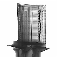
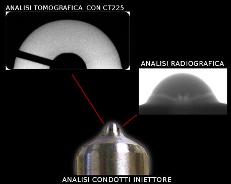
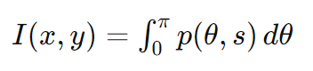
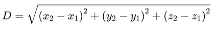
 " Tomographic images, Automotive, valve part, composite, Electronic"Industrial tomography revolutionizes the non-destructive inspection process, allowing operators to "peek" inside objects without causing any damage.With its advanced imaging techniques and applications across various industrial sectors, this technology provides a detailed and accurate view of the internal structure of objects, contributing to the quality, safety, and performance of industrial products.ConclusionsIndustrial tomography, with its approach grounded in solid physical principles and enriched by the use of mathematical formulas for image reconstruction, stands at the forefront of material engineering and non-destructive analysis.Its applications span multiple industries, offering a powerful tool for innovation and the continuous improvement of production processes and product quality.Chapter 2: "Crossing the Veil: Fundamentals of Metrological Tomography"Here, we will explore the concepts behind metrological tomography, a specialized branch that combines tomography with metrology for accurate measurement of components and structures.We will see how this technique overcomes the limitations of traditional measurements, offering a detailed and precise view.Metrological Tomography: Innovation in the Union of Tomography and MetrologyMetrological tomography represents a technological forefront that integrates the precision of metrology with the three-dimensional imaging capabilities of tomography, providing advanced solutions for the detailed analysis of components and internal structures.This approach not only surpasses the limitations of traditional measurement techniques but also opens new possibilities for inspection and dimensional verification across various fields, from industrial production to material engineering.Principles and ApplicationsMetrological tomography utilizes the principle of differential radiation absorption to generate detailed images of objects. X-rays, for example, are absorbed differently by various materials depending on their density and chemical composition.When X-rays pass through an object and are detected by a detector, the intensity of the absorbed rays varies, creating a contrast that can be transformed into 3D images of the object's interior.Practical ExampleConsider a complex mechanical component, such as an injector for internal combustion engines.The precision of its internal microgeometries, not directly accessible through conventional inspection methods, is crucial for its functionality.Using metrological tomography, it is possible not only to visualize but also to precisely measure the internal dimensions, such as the injection channels or combustion chambers, without altering or destroying the component." Tomographic analysis of injector ducts"The Basic Formula for Image ReconstructionComputational tomography utilizes mathematical algorithms to reconstruct three-dimensional images from radiographic projections collected at different angles.An example of a formula used in this process is the Filtered Back Projection (FBP) Reconstruction Algorithm, which can be expressed as:where I(x,y) represents the intensity of the point in the reconstructed image at coordinates (x,y), p(θ,s) is the radiographic projection at an angle θ and a distance s from the rotation center, and the integral is calculated over all projection angles from 0 to π radians.Advantages and ChallengesThe use of metrological tomography offers numerous advantages, including the ability to inspect the completeness of complex assemblies, identify internal defects not visible externally, and measure internal dimensions with precision unthinkable with traditional methods.However, challenges related to data processing and image interpretation require specialized skills and advanced software solutions to maximize the effectiveness of this technology.In conclusion, metrological tomography opens new horizons in the measurement and analysis of components and structures, providing valuable tools to improve the quality and reliability of products across various industrial sectors.Navigating Beyond Traditional Limitations: Comparison between Standard and Metrological Industrial TomographyThe challenge of accurately measuring objects of varying complexity, especially when they present intricate geometries or hidden internal parts, has always been a critical point in industries requiring high precision and quality.While traditional measurements encounter these limits, two advanced approaches of tomography emerge: standard industrial tomography and metrological tomography.Both offer solutions for inspecting the interior of objects, but with notably different purposes and levels of precision.Standard Industrial TomographyStandard industrial tomography utilizes radiation, such as X-rays or gamma rays, to penetrate objects and create images of their internal structure.The fundamental principle is the differential absorption of radiation, which varies according to the density and composition of materials.This method is extremely useful for identifying internal defects like cracks, voids, or inclusions in materials without destroying the object.Practical ExampleFor instance, a casting component made of aluminum can be examined to detect porosity or non-metallic inclusions within it.Standard industrial tomography can reveal these imperfections, providing a three-dimensional image that locates the defect within the piece."Three-dimensional internal tomographic image"Industrial Metrological TomographyBeyond the simple identification of defects, metrological tomography goes further, combining imaging techniques with highly precise measurement methods.It uses sophisticated image processing algorithms and advanced calibration systems, not just to visualize but also to accurately measure the dimensions and shapes of objects.This approach is crucial in contexts where dimensional specifications and geometric conformity are essential for the functionality and performance of the product.Consider wanting to measure the internal diameter of a small mechanical component with complex geometries.Metrological tomography employs the concept of 3D volumetric reconstruction, where the dimension D of an internal detail can be calculated through the equation:where (x1,y1,z1) and (x2,y2,z2) represent the three-dimensional coordinates of the opposite points of the measured detail.This precision is crucial for applications such as the dimensional analysis of components in high-technology sectors, where every micrometer counts."Tomographic image with measurements"ConclusionWhile standard industrial tomography provides a powerful solution for identifying internal defects without destroying the object, metrological tomography elevates inspection capabilities to a higher level.By offering not only visualization but also precise measurements of the dimensions and shapes of objects, it ensures the fulfillment of the stringent quality and performance standards required in sectors such as aerospace, automotive, and medical.This distinguishes metrological tomography as a fundamental pillar for quality assurance and the optimization of production processes across a wide range of industrial applications.Chapter 3: Exploring Different Tomography Systems: X-Ray Tubes and DetectorsIn the dynamic field of industrial and metrological tomography, the relentless push towards optimizing resolution and precision has led to the development of advanced X-ray beam focusing technologies.These technologies, specifically systems with Linear Accelerator, Mini-focus, Micro-focus, and Nano-focus, represent the frontier of innovation, offering unprecedented solutions for the detailed analysis of a wide range of materials and components.In this chapter, we will delve into an in-depth examination of these systems, outlining how each contributes to elevating tomography to unprecedented levels of detail and accuracy."Comet-Yxlon's Microfocus and Nanofocus Tubes"Linear Accelerators: Penetration and Precision to New LevelsLinear accelerators (Linacs) are devices capable of accelerating electrons to speeds close to that of light, generating high-energy X-rays when these electrons strike a metal target.The ability to produce high-energy beams translates into superior material penetration, making these systems ideal for inspecting large thickness and high-density components, such as those used in the aerospace industry, nuclear energy, and defense applications.The uniqueness of linear accelerators lies in their flexibility and precision, offering operators the ability to adjust the beam energy to optimize image quality based on the specific characteristics of the sample analyzed.This versatility allows for high-resolution imaging of complex objects while maintaining excellent material discrimination and detailed visualization of internal structures.X-ray linear accelerators (LINACs) also find application in the industrial field, where they are used for a variety of purposes, including non-destructive testing, sterilization, and material treatment.Here are some key data and features of X-ray linear accelerators in industrial use:
- Non-Destructive Testing (NDT): LINACs are used in non-destructive testing to inspect components and structures without damaging them. This is particularly useful in sectors such as aerospace, automotive, and oil and gas, where ensuring the structural integrity of materials and components is critical. Linear accelerators generate X-rays or electron beams that can penetrate materials and reveal internal defects such as cracks, porosity, or foreign inclusions.
- Sterilization: Linear accelerators are used for the sterilization of medical devices, food packaging, and sometimes food itself. The high-energy electron beam kills contaminating microorganisms by altering their DNA. This method is quick, effective, and leaves no chemical residues, making it a preferable sterilization technique for many products.
- Material Treatment: In material treatment, LINACs can be used to alter the chemical, physical, and mechanical properties of materials. For example, they can be employed to cross-link polymers, improving their heat resistance and mechanical stress resilience, or to surface-harden metals.
- Security and Inspection: Linear accelerators are used in scanners for container and vehicle inspection at ports and borders. These systems can penetrate dense loads, providing high-resolution images for the inspection of goods, revealing contraband, illicit substances, or hidden explosive devices.
- Research and Development: In R&D, LINACs are used to study materials at the molecular and atomic level. Applications include X-ray crystallography, which helps determine the structure of crystals, and high-energy radiography to explore material properties under stress or in extreme conditions.
- Energy and Power: Industrial linear accelerators vary greatly in terms of energy and power, from a few MeV to tens of MeV, depending on the application. For example, sterilization and material treatment may require lower energies, while non-destructive testing of thick or dense components may need higher energies.
- Costs and Maintenance: Although purchasing and maintaining an industrial LINAC can represent a significant investment, the efficiency and effectiveness of these systems can lead to long-term savings by reducing production costs, improving product quality, and reducing the rate of scrap.
In conclusion, X-ray linear accelerators play a crucial role in various industrial applications, from security to product quality to research.Their ability to provide non-destructive inspections, advanced material treatments, and effective sterilization makes them valuable tools in multiple industrial sectors."Siemens' 15MeV Linear Accelerator"Mini-focus: A Closer LookThe mini-focus system is distinguished by its ability to concentrate the X-ray beam on a relatively small point of the sample.This refined focus significantly improves spatial resolution and detail definition, making it possible to reveal extremely fine structures and conduct in-depth analysis of samples with a level of detail previously unimaginable.Applications requiring supreme precision, such as the analysis of high-density materials or meticulous characterization of components on a microscopic scale, find in mini-focus an unparalleled ally.Mini-focus X-ray tubes are used in various industrial sectors, especially for non-destructive testing (NDT), material analysis, and electron microscopy.These tubes are designed to generate a very small focal point, which improves the resolution of X-ray images, allowing the identification of very fine defects or detailed analysis of materials.Here are some key data and features relevant to mini-focus X-ray tubes in the industrial field:
- Focal Spot Size: Mini-focus X-ray tubes generally have a focal spot ranging from less than 200 micrometers (µm) to a few millimeters. This allows for high-resolution images, essential for the inspection of small components or with high precision requirements.
- Applications: They are widely used in industries such as electronics, aerospace, automotive, and precision mechanics. For example, in the electronics industry, mini-focus X-ray tubes are used to inspect welds, connectors, and internal components of printed circuit boards. In aerospace, they contribute to the inspection of composite materials and metal parts to reveal internal defects such as cracks, porosity, or inclusions.
- Technology: Mini-focus X-ray tubes can operate at both low and high energy, depending on the application. Low-energy systems are typically used for lighter and thinner materials, while high-energy systems are required to penetrate thicker and denser materials.
- Advantages: Compared to conventional X-ray tubes, mini-focus offers better image resolution, allowing for more detailed and precise material analysis. This is particularly useful for identifying very small defects that might not be detected with larger focal systems.
- Micro-CT: One of the significant applications of mini-focus X-ray tubes is micro-computed tomography (micro-CT), which allows 3D visualization of small objects with high-resolution details. This is useful not only for quality control but also for research and development, allowing internal analysis of samples without destroying them.
- Limitations: While the advantages of mini-focus technology are clear, it requires more expensive equipment and operations that are sometimes more complex compared to traditional X-ray systems. The management and maintenance of such systems require qualified personnel and specific procedures.
- Future Developments: Ongoing research in the field of X-ray tubes aims to further improve resolution, reduce focal spot sizes, and increase the power and efficiency of tubes. This will allow for even broader and more detailed applications across various industrial sectors.
In conclusion, mini-focus X-ray tubes represent a key technology for precision inspection and analysis in multiple industrial sectors.Their ability to provide high-resolution images significantly improves the quality of control and material analysis, thus contributing to innovation and product reliability."Comet closed Minifocus X-ray tubes"Micro-focus: Beyond the Boundaries of the MinuteDirectly evolving from mini-focus, the micro-focus system embodies a further qualitative leap in X-ray beam focusing, achieving an even smaller focal point.This capability for extreme concentration translates into superior spatial resolution and the ability to discern far finer details within the sample.Applications benefiting from this technology are those requiring resolution at the edge of the conceivable, such as the analysis of nanostructured materials or the search for microscopic defects, offering an unprecedented view of the invisible world.Micro-focus X-ray tubes are advanced tools for precision imaging, widely used in various industrial applications for non-destructive testing (NDT), material analysis, electron microscopy, and more.These tubes are designed to generate a very small focal point, significantly improving image resolution compared to traditional X-ray systems. Without referring to specific manufacturers, here are some key aspects of micro-focus X-ray tubes in the industrial context:


"Comet-Yxlon's high-power microfocus at 225keV"Nano-focus: Mastering the Infinitely SmallThe nano-focus system represents the pinnacle of innovation in X-ray beam focusing, enabling the achievement of nanometric dimensions at the point of contact with the sample.This extreme degree of precision unlocks the door to the highest conceivable spatial resolution, allowing for the exploration of previously unreachable details.Applications leveraging this technology range from the analysis of nanostructured materials to the characterization of nanoparticles, setting new standards for precision and detail.Nano-focus X-ray tubes used in the industrial field are advanced tools that provide high-resolution images for quality control, non-destructive testing (NDT), and other applications requiring fine details and precision.These tubes are particularly valuable in sectors such as aerospace, automotive, electronics, and precision mechanics. Here are some features and relevant data without referring to specific brands:

 "Comet-Yxlon's high-power nanofocus at 190KeV"The choice of the most suitable focusing system varies depending on the specific requirements of the application in question.Each of these systems offers a unique set of advantages and limitations.However, regardless of the choice, the continuous evolution of industrial and metrological tomographic technology is constantly expanding the boundaries of what can be analyzed and understood, ushering in new eras of discovery in the field of materials science and beyond."Table of different systems with focal spot size and penetration"Detectors in Computed Tomography Systems: Innovations and PerformanceIn modern Computed Tomography (CT) systems, the evolution of detectors plays a crucial role in enhancing performance and image quality.Today, these systems are commonly equipped with Digital Detector Arrays (DDA) or Linear Detector Arrays (LDA), each with specific features and applications that meet the growing demands of the industry.Digital Detector Arrays (DDA): Precision and ClarityDDAs represent the forefront in replacing traditional radiographic films and Computer Radiography (CR) systems, thanks to their superior sensitivity, resolution, and bit depth.These modern detectors offer exceptionally clear images and high contrast, a breakthrough for diagnostic accuracy.The flat surface of the detector and the geometry of square pixels effectively eliminate image distortions, ensuring unprecedented visual fidelity.With pixel sizes ranging approximately from 50 µm to 400 µm, DDAs can support a range of image acquisition frequencies from about 2 frames per second (fps) up to 100 fps, with the ability to adjust sensitivity through various levels of amplification, thus optimizing image quality based on specific diagnostic requirements.For additional information, follow the link: DDA."Flat Panel and Linear Array Modular system by Comet-Yxlon"Linear Detectors (LDA): Specialization for Precision ScanningLDA stand out for their specialized applications in fan beam CT scans, particularly suitable for analyzing components with considerable thickness.These detectors offer pixel resolution ranging from approximately 80 µm to 800 µm, allowing detailed imaging even of the densest structures.A notable aspect of some LDA models is their ability to adjust temperature, ensuring consistently high performance and uncompromised stability even under intense usage conditions.A significant advantage of these systems is their detector modularity, allowing for the replacement of individual modules if necessary, thus reducing costs and maintenance times.With image update frequencies ranging from approximately 30 fps to 600 fps, LDAs meet the requirements of rapid acquisitions without sacrificing image quality. For additional information, follow the link: LDA."Linear Array system FF50 by Comet-Yxlon"ConclusionContinuous innovation in the field of detectors for Computed Tomography is revolutionizing the diagnostic and analytical capabilities of this technology.Whether it's DDA, with their excellent resolution and contrast for general applications, or LDA, specialized for precision scans on dense components, these technologies are setting new standards of quality and precision in imaging.The adaptability, precision, and efficiency of modern detectors significantly expand the potential of computed tomography, promising increasingly significant advancements in the field of industrial imaging."Micro-Tomography and its interaction with other sectors"Chapter 4: "Beyond the Image: Processing and Analysis of Tomographic Data"Segmentation and image analysis constitute two fundamental phases in the image processing process, particularly in applications ranging from medical engineering to materials analysis.These advanced processes allow for the isolation and examination of specific structures or regions of interest (ROI) within an image, transforming raw data into valuable and actionable information."Tomography - Image analysis with VG"In-depth Analysis TechniquesAfter successfully segmenting the regions of interest using one of the aforementioned techniques, image analysis becomes the focal point for translating this visual data into actionable knowledge.Comprehensive analysis encompasses not only the measurement of area and volume but extends its scope to more complex parameters such as surface roughness, grain orientation, or spatial correlation between different material phases."Tomography image analysis with various applications"Image Segmentation: Techniques and ApplicationsImage segmentation is the process of dividing an image into parts or regions that have a closer meaning compared to the entire image.These regions can represent individual objects or areas of specific interest within the image. Segmentation techniques vary in complexity and adapt to different types of images and analysis requirements.Main TechniquesContour-Based Segmentation
- Threshold-Based Segmentation: This technique involves dividing the image into regions based on variations in pixel intensity. A threshold T is applied to separate the pixels of interest from the background or other objects.


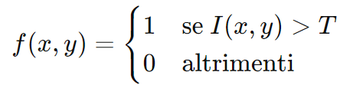
Where I(x,y) represents the intensity of the pixel and f(x,y) represents the result of segmentation.
2. Contour-Based Segmentation: It uses contrast differences to detect object edges. Techniques like the Canny algorithm identify boundaries through intensity gradients, highlighting the edges of objects.3. Region-Based Segmentation: This method groups adjacent pixels or voxels with similar properties, such as texture or color, to form homogeneous regions. Algorithms like region growing and split and merge are practical examples of this technique.
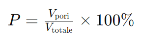
Practical ExampleLet's consider the analysis of a medical image, such as an MRI scan of the brain.Using threshold-based segmentation, it is possible to isolate specific brain structures based on their signal intensity, facilitating studies on the volume and morphology of brain tissue.For example, distinguishing white matter from gray matter or identifying areas of abnormality.Image Analysis: Information ExtractionOnce segmentation is completed, image analysis allows for the extraction of quantitative and qualitative information from segmented regions.This may include calculating areas, volumes, object shapes, texture, and other relevant metrics."Histogram table of defects"Connectivity and Porosity AnalysisIn porous samples or foamy materials, determining pore connectivity or measuring pore size distribution can be of fundamental importance. For instance, the total porosity of a material can be calculated as the ratio between the pore volume and the total volume of the sample:Where P is the percentage porosity, Vpores is the volume of pores identified through segmentation, and Vtotal is the total volume of the sample." Image of porosity inside a cast component."Calculation of Area and Volume: They are computed by summing the pixels or voxels within the segmented region. For an image with known resolution, the volume V of an object can be calculated as:


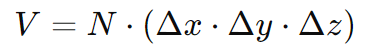
Where N is the number of voxels in the segmented object, and Δx, Δy, and Δz represent the physical dimensions of each voxel."Image showing porosity and misalignment of engine cylinder heads"

Texture CharacterizationTexture analysis provides a more detailed view of the spatial arrangement of pixel or voxel intensities, giving insights into patterns, directionality, or regularity present in the image.Statistical methods, such as Gray-Level Co-occurrence Matrix (GLCM), can quantify texture by examining the frequency of pairs of pixels with specific intensity values and orientations.
Example ApplicationIn the field of materials engineering, image segmentation and analysis can be employed to quantify the distribution of phases or the presence of defects in a metallographic sample.Calculating the percentage of a specific metallic phase within.
Vision of the different phases of tomography a. Real Component, b. Image Acquisition,c. Point Cloud Volume, d. Virtual Component Image, e. Component AnalysisAdvanced Application Examples
- Biomedical Engineering: Image segmentation and analysis play a key role in disease diagnosis and monitoring.

For instance, segmenting MRI brain images can help quantify the progression of neurodegenerative diseases by measuring the volume reduction of specific brain structures.
"Part of a Rhinoceros Head"

2. Environmental Research:In the analysis of satellite images, segmentation can be used to distinguish different land covers such as water bodies, forests, or urban areas, enabling studies on land use or climate change.
"Viewing and Analysis of a Rock Sample"

3. Quality Control in Manufacturing Industry:Image analysis of industrial components can detect production defects, such as cracks or misalignments, ensuring that only parts that meet rigorous quality criteria reach the market.
"Internal Inspection of Electronic Components"In conclusion, image segmentation and analysis offer powerful tools to transform images into understandable and actionable insights.Through the application of advanced techniques and detailed understanding of the involved processes, significant value can be extracted from images in a variety of fields, enhancing research, production, and diagnosis in previously unimaginable ways.Chapter 5: "Industrial Applications: From Automotive to Electronic Components"Here, we will examine a wide range of industrial applications of tomography, from inspecting automotive components to assessing the quality of electronic devices.We will discover how this technology integrates into manufacturing processes to improve product quality and reliability.Industrial Applications: From Automotive to Electronic ComponentsIndustrial tomography has revolutionized non-destructive inspection across various industrial sectors, offering detailed insight into the internal structure of objects without damaging them.In this chapter, we will explore the numerous applications of this technology, from inspecting automotive components to evaluating the quality of electronic devices.We will uncover how tomography integrates into manufacturing processes to enhance product quality and reliability. "Application of Tomography in Electronics: Inspection of Integrated Circuits and Ball Grid Arrays (BGA)"Automotive Component InspectionIn the automotive industry, tomography finds fundamental applications in inspecting critical components such as engines, transmissions, and suspension systems.This technology enables the detection of internal defects such as porosity in castings, foreign material inclusions, or hidden fractures that could compromise the performance and safety of vehicles.Additionally, tomography is employed to assess component compliance with design specifications and to optimize production processes."Image showing porosity and deviation of an automotive component"Evaluation of Electronic Devices QualityIn the field of electronic components, tomography is widely used for inspecting printed circuit boards, microelectronic components, and assembly devices.This technology enables the detection of defects such as faulty solder joints, missing contacts, or chip damage that could compromise the functionality of electronic devices.Additionally, tomography is utilized to conduct reliability and longevity analysis of products, contributing to ensuring the quality and reliability of electronic devices."Porosity image in BGA soldering elements"Integration into Production ProcessesTomography is increasingly tightly integrated into production processes, becoming a crucial phase in quality control and process optimization.Thanks to its ability to provide detailed information about the internal structure of objects, this technology enables the timely identification and correction of any defects or anomalies, reducing waste and improving overall production efficiency.Furthermore, tomography supports the development of new materials and technologies, contributing to innovation and the competitiveness of companies.In conclusion, industrial tomography emerges as a versatile and powerful technology for non-destructive inspection across a wide range of industrial sectors.From automotive to electronics manufacturing, this technology integrates into production processes to enhance the quality, reliability, and safety of products, contributing to industry advancement and customer satisfaction.Chapter 6: "Navigating Anomalies: Defect Detection and Analysis"In this chapter, we will delve into the crucial role of tomography in detecting and analyzing defects and anomalies in materials and products.We will explore advanced techniques for identifying and characterizing defects of various types, contributing to the safety and reliability of manufactured goods.Navigating Anomalies: Defect Detection and AnalysisIn the realm of non-destructive inspection, the ability to detect and analyze defects and anomalies in materials and products plays a crucial role in ensuring their safety and reliability.In this chapter, we will explore the fundamental role of tomography in detecting and analyzing defects of various types, not only identifying them but also characterizing them to fully understand their implications and potential risks.Defect IdentificationIndustrial tomography provides an effective solution for defect identification, thanks to its ability to provide high-resolution three-dimensional images of the internal structure of objects.Through sophisticated image processing algorithms, defects such as porosity, foreign material inclusions, fractures, or material structure discontinuities can be identified.This technology accurately visualizes the position, shape, and dimensions of defects, providing crucial information for evaluating the quality and integrity of materials and products."Porosity visualization and dimensional indication through color coding"Defect CharacterizationIn addition to identification, tomography also allows for the characterization of defects, i.e., understanding their characteristics and causes.This process involves detailed analysis of tomographic images to assess the nature, extent, and impact of defects on the performance and safety of manufactured goods.For example, it is possible to determine whether a defect is caused by a faulty manufacturing process, non-compliant materials, or environmental factors, in order to take the necessary corrective and preventive measures."Dimensional inspection of components using tomography"Advanced Analysis TechniquesIndustrial tomography offers a wide range of advanced analysis techniques for thoroughly characterizing defects.These include segmentation and quantification of defect characteristics, assessment of their distribution and morphology, and analysis of underlying causes through statistical analysis and computational modeling.These techniques enable a comprehensive understanding of defects, providing a solid foundation for optimizing manufacturing processes and improving product quality.In conclusion, industrial tomography plays a crucial role in detecting and analyzing defects and anomalies in materials and products.Thanks to its ability to accurately and reliably identify and characterize defects, this technology contributes to the safety, reliability, and quality of manufactured goods, playing a fundamental role in ensuring compliance with production standards and maintaining consumer confidence.Chapter 7: "Enhancing Precision: Precision Metrology with Tomography"In this chapter, we will analyze how metrological tomography enables the measurement of components with high precision and accuracy.We will explore the challenges and solutions in high-precision metrology and how tomography contributes to overcoming them.Enhancing Precision: Precision Metrology with TomographyPrecision metrology is fundamental to ensuring the quality and performance of industrial products.In this chapter, we will examine how metrological tomography allows for the measurement of components with high precision and accuracy.We will explore the challenges and solutions in high-precision metrology and how tomography contributes to overcoming them.Precision Metrology and Associated ChallengesPrecision metrology faces several challenges, including the geometric complexity of components, the need for non-invasive measurements, and the requirement for high accuracy in measurements.Additionally, the presence of complex internal structures or multi-component materials can further complicate the measurement process.Addressing these challenges requires the adoption of advanced techniques and instrumentation that can ensure reliable and reproducible measurements.Metrological Tomography: An Advanced SolutionMetrological tomography emerges as an advanced solution for precision metrology, enabling accurate measurement of complex components and internal structures.This technique combines the principles of tomography, which provides detailed three-dimensional images, with those of metrology, which deals with the measurement of dimensions and shapes of objects.Thanks to its ability to visualize and measure internal details, metrological tomography overcomes the limitations of traditional measurement techniques, allowing for accurate assessment of components even with complex geometries.Challenges Overcome by Metrological TomographyMetrological tomography successfully addresses many of the challenges associated with precision metrology.Its ability to provide high-resolution three-dimensional images allows for precise evaluation of the dimensions, shapes, and characteristics of objects, including the detection of internal defects or small dimensional variations.Furthermore, metrological tomography enables non-invasive measurements, allowing for detailed analysis without damaging the inspected components.Contribution to Quality and InnovationThanks to its ability to improve the precision and accuracy of measurements, metrological tomography contributes to quality and innovation in manufacturing processes.This technology enables the optimization of component design and production, reducing errors and improving the reliability of final products.Additionally, metrological tomography supports the development of new technologies and materials, contributing to the continuous evolution of the industrial sector.In conclusion, metrological tomography represents an advanced solution for precision metrology, enabling accurate measurement of complex components and internal structures.By successfully addressing the challenges of high-precision metrology, this technology contributes to quality, reliability, and innovation in manufacturing processes, playing a fundamental role in ensuring compliance with quality standards and promoting technological development.Chapter 8: "Metrological Tomography vs. Traditional Tomography: Key Differences in Application and Purpose"Differentiating between Metrological and Non-Metrological Tomography: In-depth Analysis of their Features and ApplicationsThe main distinction between a metrological tomograph and a non-metrological one is intrinsic to their purpose and scope of use. Let's explore these differences in more detail:Non-Metrological Tomography:The non-metrological tomograph, as the name suggests, is designed for purposes beyond metrology, i.e., the science of measurements.This type of tomography is commonly used to examine and analyze the internal structure of objects without the primary goal of providing precise dimensional measurements.Instead, its use focuses primarily on defect detection, material analysis, and three-dimensional visualization of objects without the need for specific dimensional measurements.The applicative flexibility of non-metrological tomographs makes them valuable tools in a wide range of sectors and applications.From scientific research and engineering to archaeology and biological sciences, these devices are used to explore the internal structure of objects of various sizes and compositions, providing a detailed insight into their morphology and composition without the need for precise dimensional measurements.While non-metrological tomographs are capable of detecting defects and anomalies in the structure of objects, their main goal often lies in analyzing the internal structure for purposes of research, scientific exploration, or material analysis.In summary, the main distinction between a metrological tomograph and a non-metrological one lies in their primary intent and specific application.While the former is aimed at providing precise measurements of the dimensions and shapes of objects, the latter focuses more on the visualization and analysis of internal structure without necessarily concentrating on dimensional accuracy."Non-Metrological Tomography Facility FF35"Key Features of Metrological Tomography:Metrological tomography, an advanced form of tomography primarily used for measurement and metrology purposes, offers a range of distinctive features that make it a fundamental technology in a wide array of industrial sectors.One of the most relevant aspects of metrological tomography is its ability to provide extremely precise measurements of the dimensions and shapes of the objects under examination.Through the use of sophisticated image processing algorithms and advanced calibration systems, measurements with precision up to fractions of a micron can be achieved.This level of precision is crucial, especially in sectors where even the smallest deviations from specifications can have significant consequences on product quality and performance.Another distinctive feature of metrological tomography is its ability to acquire images with high spatial resolution. By utilizing optical systems and highly sensitive sensors, metrological tomographs can capture structural details down to microscopic levels."Metrological Tomography Facility FF35"This high level of detail allows for the identification and measurement of even the smallest features and discontinuities of objects, contributing to ensuring an accurate assessment of their dimensions and shapes.Before being used for metrological purposes, metrological tomographs must undergo rigorous calibration and verification procedures.These operations are essential to ensure that the measurements obtained comply with international metrology standards and that the system is accurately calibrated to provide reliable and repeatable results over time.Regular calibration and systematic verification are therefore essential to maintain the reliability and accuracy of metrological results over time.Thanks to its flexibility and ability to measure objects of various sizes, geometries, and materials, metrological tomography finds application in a wide range of industrial sectors.It is successfully employed in the automotive, aerospace, medical device manufacturing, and many other sectors where dimensional and geometric precision is critical to ensuring product quality and performance.Its ability to adapt to different application contexts makes it a valuable resource for the needs of advanced metrology in complex and continuously changing industrial environments.Finally, metrological tomographs can be easily integrated with quality analysis and control systems to automate the inspection and measurement process.This integration allows for continuous quality control during the production process, identifying any non-conformities and enabling timely interventions to ensure compliance with quality requirements.The ability to integrate with other industrial systems and processes contributes to improving the efficiency, productivity, and consistency of quality control operations.In conclusion, metrological tomography represents a powerful technology for the precise measurement of the dimensions and shapes of objects, offering a reliable and versatile solution for the needs of advanced metrology in a wide range of industrial sectors.Thanks to its extreme precision, high spatial resolution, accurate calibration, applicative flexibility, and integration potential with quality control systems, metrological tomography proves to be an indispensable tool for ensuring the quality, reliability, and performance of industrial products.Standards and Regulations in Metrological Tomography: Guarantees of Reliability and PrecisionMetrological tomographs must adhere to a series of standards and regulations to ensure that the measurements obtained are accurate, reliable, and compliant with international metrology standards. Some of the main standards used in a metrological tomograph include:


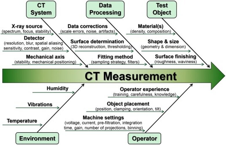
1. ISO 10360: This standard defines the requirements for the verification of coordinate measuring machines (CMMs), including X-ray tomography machines. It provides guidelines for evaluating machine performance, including accuracy, repeatability, and linearity.2. ISO 16742: This standard provides guidelines for assessing the performance of X-ray tomographs used for non-destructive testing. It focuses on defining calibration, verification, and result validation procedures obtained through tomography.3. ASTM E1695: This standard, developed by the American Society for Testing and Materials (ASTM), concerns the standard practice for verifying the performance of X-ray tomography systems. It provides guidelines for verifying spatial resolution, contrast sensitivity, and measurement accuracy.4. VDI/VDE 2630: This standard, developed by the German Association for Information Technology, Telecommunications and Electronics (VDI/VDE), concerns measurement methods for evaluating the performance of X-ray tomographs. It focuses on defining calibration, verification, and validation procedures to ensure measurement reliability.5. NISTIR 7034: This document, published by the National Institute of Standards and Technology of the United States (NIST), provides guidelines for assessing the performance of X-ray tomography systems. It focuses on defining calibration and verification procedures to ensure that the obtained results are accurate and repeatable.
These are just some of the standards used in tomographic metrology.It is important to note that the choice of standards often depends on the geographical region, industrial sector, and specific applications of the metrological tomograph.However, compliance with such standards is essential to ensure the quality and reliability of measurements obtained through metrological tomography."MPE: The Barometer of Reliability in Measurements"The term "MPE" stands for "Maximum Permissible Error."When referring to "Measuring Accuracy," it is common to use the term "MPE" to indicate how much the maximum allowable error in a measurement can deviate from the true value of the measured quantity."Measuring Accuracy" refers to the ability of a measurement system to approach the true or accepted value of a measured quantity. This concept is essential in metrology, where the precision and reliability of measurements are crucial.The "Maximum Permissible Error" (MPE) defines the maximum limit within which the measurement error can vary from the true value of the measured quantity without compromising the reliability of the measurement itself.For example, considering a metrological tomograph that provides dimensional measurements of a mechanical component, the MPE will indicate the maximum error allowed relative to the actual measurements of the component.If the MPE is, for example, ±0.05 millimeters for a particular measurement, this means that the measurement error can vary up to ±0.05 millimeters from the true value without the measurement being considered unreliable.In summary, the MPE provides a critical parameter for evaluating the precision and reliability of measurements made by a measurement system, such as a metrological tomograph.A lower MPE value indicates higher precision of the measurement system, while a higher MPE value indicates lower precision."MPE (Maximum Permissible Error) metrological verification"In a world where clarity and precision of information are paramount, especially in the manufacturing sector of measuring instruments, how the technical capabilities of a product are communicated can make a difference in perceiving its value.In the context of metrology, the science of measurements, there are often communicative challenges related to the interpretation and use of technical terms.This overview aims to shed light on how, in the fervor of marketing, some of these terms can be interpreted differently, used improperly, or even exchanged, creating fertile ground for misunderstandings and misconceptions.The crux of the matter lies in the tension between the need to communicate the qualities of a measuring instrument accurately and the desire to make this information accessible and compelling to a broad and diverse audience.In this perspective, marketing strategies play a crucial role, shaping the perception and interpretation of technical terms in a way that is both accurate and engaging.However, the challenge lies in maintaining technical integrity without overwhelming the consumer with complex details that could hinder understanding rather than facilitate it.This balance requires careful navigation, where communication clarity marries with the authenticity and precision of technical information.The manufacturing sector of measuring instruments, therefore, finds itself at a communicative crossroads: on one hand, the need to remain faithful to scientific and technical rigor, and on the other, the necessity of speaking a language that is both accurate and accessible.Overcoming these challenges would not only help build greater trust between manufacturers and consumers but also elevate the level of understanding and appreciation of the subtle dynamics that govern the world of metrology.Precision vs. Accuracy:
- Precision: Refers to the consistency of measurements, i.e., the ability of an instrument to provide results close to each other under repeated test conditions. It is important to note that a series of measurements can be precise without being accurate, meaning the results can be close to each other but far from the true or accepted value.
- Accuracy: Indicates how close a measurement is to the true or accepted value. An instrument is considered accurate if its measurements are close to the reference value, regardless of the repeatability or consistency of those measurements.
Improper Use of Terms:
- In the realm of marketing and product presentation of measuring instruments, the terms precision and uncertainty may be used interchangeably or inaccurately, generating confusion. This approach may be influenced by the positive perception associated with accuracy and the negative perception associated with uncertainty, leading to an optimistic presentation of a product's capabilities.
Accuracy Statements:
- Manufacturers often provide accuracy statements that should qualitatively describe the performance of a measuring instrument. These statements, however, may include quantitative values aimed at depicting the measurement capabilities of the instrument across a wide variety of conditions, although there is a trend towards standardization to make these statements more representative of actual performance.
Maximum Permissible Error (MPE):
- MPE is a key concept in defining accuracy, establishing an upper limit to the acceptable deviation for a measurement from a reference value. This limit helps ensure that the instrument is reliable for a specific measurement task, defining the maximum tolerance beyond which the precision of an instrument is considered insufficient.
Traceability and Uncertainty:
- Traceability refers to the ability to link measurements made by an instrument to national or international standards, ensuring that the results are universally recognized. Measurement uncertainty is a fundamental component of this process, providing an estimate of the expected variability around the result of a measurement. By defining an extended uncertainty, limited by the MPE, manufacturers can establish a level of confidence in the measurement capabilities of an instrument, ensuring that it is suitable for the intended purpose.
Through a comprehensive understanding of these concepts, it is possible to more accurately assess the performance of measuring instruments and the reliability of the results obtained, facilitating the selection of the most suitable tools for specific needs.Chapter 9: "Challenges and Solutions: Optimization of Tomographic Processes"In this chapter, we will address practical challenges in achieving efficient and accurate tomographic scans.We will explore best practices and innovative solutions to optimize data acquisition and analysis processes.Challenges and Solutions: Optimization of Tomographic ProcessesWithin the realm of tomographic scans, it is essential to address practical challenges to ensure that data acquisition and analysis processes are efficient and accurate.In this chapter, we will explore the key challenges and innovative solutions to optimize tomographic processes, enabling high-quality results to be obtained more quickly and efficiently.Practical Challenges in Tomographic ScanningPractical challenges in tomographic scanning may include the geometric complexity of the objects to be examined, the need to balance spatial resolution with scanning time, and the management of large volumes of data generated during the process.Additionally, factors such as noise reduction in images, distortion correction, and artifact minimization can influence the overall quality of the results obtained.Best Practices for Optimizing Tomographic ProcessesTo address these challenges, it is important to adopt best practices to optimize tomographic processes.This may include proper selection of scanning parameters, such as X-ray source voltage and current, exposure time, and angular resolution.Furthermore, employing advanced filtering and image processing techniques can help improve the quality of scans and reduce unwanted noise and artifacts. "Tomography using Comet-Yxlon's Scatterfix program"Innovative Solutions to Improve Efficiency and AccuracyTechnological innovation continues to offer innovative solutions to enhance the efficiency and accuracy of tomographic processes.These may include the development of new image reconstruction techniques, the integration of artificial intelligence algorithms for automatic distortion correction and noise reduction, and the use of advanced hardware to reduce scanning times and optimize image quality.Benefits of Optimizing Tomographic ProcessesOptimizing tomographic processes brings numerous benefits, including increased operational efficiency, improved image quality, and greater reliability of the results obtained.This allows for reduced data acquisition and analysis times, enhancing overall productivity and enabling faster decision-making.Additionally, optimizing tomographic processes helps ensure the quality and reliability of the information obtained, playing a crucial role in supporting critical decisions in industrial and scientific sectors.In conclusion, addressing practical challenges in tomographic scanning requires the adoption of innovative solutions and the implementation of best practices.Optimizing tomographic processes not only enhances measurement efficiency and accuracy but also brings numerous benefits in terms of productivity, quality, and reliability of the results obtained."Comparison of image enhancements using different techniques"Chapter 10: "Looking to the Future: Trends and Developments in the Tomography Industry"Here we will examine emerging trends and technological developments in the field of industrial and metrological tomography. We will explore the latest innovations and how they are shaping the future of this discipline.Looking to the Future: Trends and Developments in the Tomography IndustryThe tomography industry continues to evolve rapidly, driven by technological innovation and the growing demand for advanced solutions for non-destructive inspection and precision metrology.In this chapter, we will examine emerging trends and technological developments in the field of industrial and metrological tomography, discovering the latest innovations and how they are shaping the future of this discipline.Advanced Imaging Acquisition TechnologiesOne of the main trends in the tomography industry involves the development of advanced imaging acquisition technologies.These include high-resolution detection systems, more powerful X-ray sources, and sophisticated image processing algorithms.These technologies enable the capture of high-quality images with increasing resolution, allowing for a more detailed assessment of the internal structure of objects.Integration of Tomography with Artificial Intelligence and Machine LearningAnother significant trend is the integration of tomography with artificial intelligence (AI) and machine learning.These technologies enable the development of advanced algorithms for automated analysis of tomographic images, improving the efficiency and accuracy of measurements.For example, AI can be used to automatically detect defects or anomalies in images, speeding up the inspection process and reducing the risk of human error.The Dragonfly platform positions itself as an advanced solution in the realm of scientific image processing, embracing the power of artificial intelligence (AI) and machine learning (ML) to maximize its already remarkable capabilities.This software, integrated into Comet-Yxlon facilities, offers an enhanced imaging system capable of analyzing complex 2D, 3D, and 4D data.The combination of AI and ML optimizes image processing, segmentation accuracy, and workflow automation, transforming Dragonfly into an even more effective tool for quantitative analysis and imaging data management.Users benefit from a user-friendly interface and advanced features such as CT reconstruction, segmentation, and hyperspectral analysis, all enriched by the sophisticated integration of intelligent algorithms, making Dragonfly a crucial ally in research and industrial applications.
 "Utilization of the Dragonfly software program used by Comet-Yxlon"Miniaturization and Portability of Tomographic DevicesAn important trend is the miniaturization and portability of tomographic devices.This allows tomography to be used in environments and applications that require flexibility and mobility, such as on-site quality control or inspection of large components.Thanks to advancements in sensor technology and device design, it is possible to create compact and lightweight tomographic systems without compromising the quality of the images.Specialized Applications and New Usage SectorsFinally, there is an increasing adoption of tomography in specialized sectors and applications.This includes the use of metrological tomography in additive manufacturing and the production of complex components, as well as the application of industrial tomography in materials analysis for scientific research and the development of new technologies.This diversification of applications contributes to the growth and diversification of the tomography industry.In conclusion, trends and developments in the tomography industry indicate a promising future full of opportunities.With increasingly advanced technologies, integration with artificial intelligence, device miniaturization, and the expansion of specialized applications, tomography will continue to play a crucial role in evaluating the quality, safety, and performance of industrial products, contributing to progress and innovation in numerous sectors.Chapter 11: "Ethical Nuances: Considerations on the Use of Industrial Tomography"The widespread adoption of industrial tomography has highlighted a range of ethical considerations that require careful reflection.In this chapter, we will delve into the ethical implications associated with the application of industrial tomography, addressing issues such as privacy protection, operational safety, and environmental impact, emphasizing the need for mindful management of this technology.Respect for Privacy and Data ProtectionOne of the primary ethical issues related to industrial tomography is respecting privacy and safeguarding data, both personal and proprietary.The technology's ability to provide detailed visualization of the internal structure of objects poses the risk of inadvertent disclosure of sensitive information.It is imperative to implement adequate security measures to ensure the confidentiality of information during inspection phases and data management.Worker Safety and HealthWorker safety and health represent a significant ethical aspect in the use of industrial tomography.Since the technology utilizes ionizing radiation for image capture, adopting stringent safety protocols to protect operators and reduce radiation exposure risks is fundamental.This entails comprehensive training, the use of personal protective equipment, and the establishment of safe operating procedures.Environmental Impact and SustainabilityThe environmental footprint of industrial tomography use presents an ethical challenge.The potential generation of radioactive or chemical waste during image acquisition and analysis requires responsible and sustainable management to prevent harm to the environment and public health.It is crucial to adopt eco-friendly waste disposal practices and reduce the environmental impact of tomographic operations.Responsible Use of TechnologyIn conclusion, the use of industrial tomography demands an ethical and responsible approach that considers the implications associated with this technology.It is essential to take measures that ensure the protection of privacy, data security, worker safety and well-being, as well as the environmental sustainability of operations.Moreover, promoting an ethical culture that values transparency, accountability, and respect for fundamental human values is imperative, ensuring that the use of industrial tomography contributes to the common good and the progress of society as a whole.Chapter 12: "Integrated Methods: Combining Tomography with Other Inspection and Measurement Techniques"In the final chapter, we will examine how tomography can be integrated with other inspection and measurement techniques to achieve a more comprehensive and detailed view of the objects under examination.We will see how the integration of these methodologies can lead to more accurate and reliable results.Integrated Methods: Combining Tomography with Other Inspection and Measurement TechniquesIn the journey towards a comprehensive and detailed understanding of the objects under examination, the integration of tomography with other inspection and measurement techniques plays a crucial role.In this final chapter, we will explore how this synergy can lead to more accurate and reliable results, offering a deeper insight into the structure and characteristics of objects.Complementarity of Inspection TechniquesOne of the main reasons for integrating tomography with other inspection techniques is the complementarity of the information provided by each methodology.While tomography offers a three-dimensional view of the object and its internal structures, other techniques such as radiography, electron microscopy, or laser scanning can provide additional details on specific aspects such as chemical composition, surface, or material microstructure.Increased Precision and ReliabilityThe integration of different inspection and measurement techniques also allows for increased precision and reliability of analyses.Combined, these methodologies can offset each other's limitations and provide a more comprehensive and accurate assessment of the objects under examination.For example, the joint use of tomography and optical microscopy can allow for a more detailed characterization of material microstructures, improving the understanding of their mechanical and thermal properties.Optimization of Inspection ProcessesThe integration of tomography with other inspection and measurement techniques also contributes to the optimization of inspection processes.By leveraging the specific capabilities of each technique, it is possible to design customized inspection approaches that maximize the efficiency and effectiveness of the analysis.This can lead to reduced inspection times, increased productivity, and better quality of the obtained data.Practical ApplicationsThe practical applications of integrating tomography with other inspection techniques are numerous and varied.These include the inspection of complex mechanical components in the automotive industry, material analysis in aerospace, and the characterization of artistic artifacts in the cultural sector.In each case, the combined use of different methodologies allows for a comprehensive and accurate assessment of objects, supporting critical decisions and promoting innovation and technological development.In conclusion, the integration of tomography with other inspection and measurement techniques offers a comprehensive and multidisciplinary approach to object analysis.This synergy allows for a deeper and more accurate understanding of the structure and characteristics of objects, improving the precision, reliability, and efficiency of the conducted analyses.These twelve chapters constitute a thorough journey through industrial and metrological tomography, providing a comprehensive overview of their applications, techniques, and challenges.Whether you are a professional in the field or a curious individual in the technological world, this manual will accompany you in exploring the depths of these ever-evolving disciplines.Comet-Yxlon: Global Excellence in Metrological TomographyWith a rich history of innovation and success in the field of metrological tomography, Comet-Yxlon stands as a pillar of the industry, offering reliable and cutting-edge solutions that meet the most demanding requirements of the sector.A Legacy of Excellence: Founded on the legacy of two giants in the inspection and metrology sector, Comet and Yxlon, the company boasts a history dating back decades of experience and expertise.The merger of these two powerhouses created a unique synergy, leading to the birth of Comet-Yxlon and solidifying its position as a global leader in metrological tomography.Global Presence: With a established presence worldwide, Comet-Yxlon serves a wide spectrum of industrial sectors in every corner of the globe.From the automotive and aerospace industries to medical and research applications, Comet-Yxlon's products are trusted by companies of all sizes for their inspection and measurement needs.Commitment to Innovation: Comet-Yxlon remains at the forefront of innovation in the industry, consistently investing in research and development to anticipate market trends and introduce cutting-edge technological solutions.This constant commitment to innovation ensures that Comet-Yxlon's customers have access to the latest technologies and best practices in the field of metrological tomography.Tailored Solutions: Comet-Yxlon understands that every application has unique requirements.Therefore, it offers customized and tailored solutions to meet the specific needs of customers.From standard inspection systems to fully customized solutions, Comet-Yxlon works closely with its customers to ensure that their needs are fully met.Global Quality Standards: Comet-Yxlon is committed to providing products and services of the highest quality, ensuring compliance with the most rigorous industry standards.Each facility undergoes stringent testing and quality controls to ensure optimal performance and long-term reliability.In conclusion, Comet-Yxlon represents a benchmark in the metrological tomography industry, combining a rich history of success with unparalleled global expertise in the sector.Thanks to its global presence, commitment to continuous innovation, and impeccable quality standards, Comet-Yxlon continues to lead the industry towards new frontiers of excellence and performance.


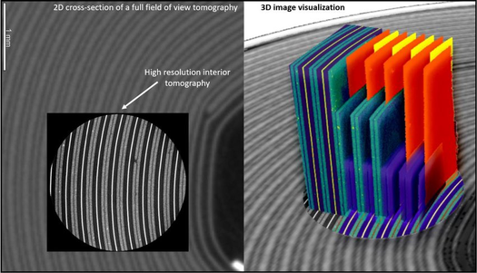

"Tomography and Micro-Tomography Systems by Comet-Yxlon"Xrayconsult is always available to provide information on this technology. To view our facilities, please click on the following link: Industrial TomographyThe information presented in this text is based on authoritative sources and can be verified through the following bibliography:
- "Industrial Tomography: A Comprehensive Introduction" by John C. Russ (2023)
- "The Fundamentals of Industrial Tomography" by Bruno P. C. Stupin (2022)
- "Introduction to Industrial Computed Tomography" by Graham S. Pettit (2021)
- "Metrological Tomography: Principles and Applications" by W. Osten, P. J. Withers, et al. (2020)
- "Advances in Tomographic Imaging for Metrology" by Xiangqian Jiang (2019)
- "Handbook of X-ray Computed Tomography for Metrology" by P. J. Withers (2018)
- "Micro-CT and Nano-CT: Principles and Applications" by Rex M. C. Luo (2022)
- "High-Resolution Computed Tomography: Principles, Techniques and Applications" by Thorsten M. Buzug (2021)
- "Recent Advances in X-ray Microtomography" by U. Bonse (2020)
- "Image Processing for Tomographic Reconstruction" by J. A. Fessler (2023)
- "Tomographic Data Analysis: Methods and Applications" by David W. Shattuck (2022)
- "Handbook of Image Processing for Biomedical Applications" by Milan Sonka, J. Michael Fitzpatrick, et al. (2021)
- "Industrial Computed Tomography: Applications in Automotive and Aerospace Engineering" by P. J. Withers (2020)
- "CT for Nondestructive Testing: Applications in Manufacturing, Aerospace and Civil Infrastructure" by T. Y. Chen (2019)
- "Electronics Manufacturing: With Advanced Materials and Processes" by Ronald Tummala (2018)
- "Defect Detection and Analysis in Industrial Computed Tomography" by J. V. Vallerga, D. W. A. Pavia, et al. (2022)
- "Non-Destructive Testing and Evaluation of Materials" by R. W. Cahn, P. K. Liaw, et al. (2021)
- "Handbook of Nondestructive Testing" by Chuck Hellier (2020)
- "Precision Metrology with Industrial Tomography" by W. Osten, P. J. Withers, et al. (2020)
- "Dimensional Metrology: Principles and Procedures" by Kenneth J. Stout (2019)
- "Advances in Manufacturing Technology and Processes" by P. K. Mishra, S. S. Mahapatra, et al. (2018)
The listed sources provide a solid foundation for the information presented and are available for detailed verification of the claims made in the text. All tomographic images in this article are from Comet-Yxlon.
